Milwaukee — Cancer diagnosis has come a long way, with noninvasive diagnostic imaging largely replacing exploratory surgery. At UWM, physicist Sarah Patch is working on the next generation of diagnostic tools: thermoacoustic imaging.
“I intentionally heat up an object a little bit, and it wants to expand because it’s heating up, so it generates an outgoing pressure pulse. We measure those pressure pulses with ultrasound receivers,” Patch explained.
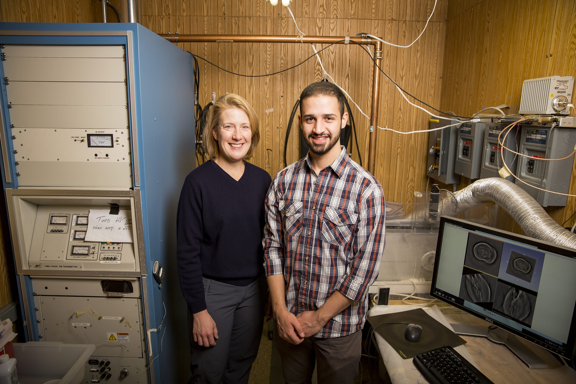
“A few years ago, we landed a grant through UWM’s instrumentation award program to purchase a research-friendly ultrasound system. Now I can use a transducer just like those in hospitals to ‘listen’ for the signal.”
Patch uses that existing technology to create 3D images of prostates with the goal of crafting a diagnostic tool that spots cancer without the risks associated with biopsies. She started by imaging cancerous prostates immediately after they were removed from patients at Froedtert & the Medical College of Wisconsin.
“Lots of folks at MCW have been very supportive of this project, from Dr. (William) See in urology, to the MCW Tissue Bank and MCW’s Clinical & Translational Science Institute,” Patch said.
To create thermoacoustic images, Patch needed a way to heat prostates uniformly. In the basement of the UWM Physics building, a souped-up FM radio transmitter propagates high-power VHF (very high frequency) pulses through her bench-top imaging system. The signal is driven by electrical conductivity. Healthy prostate glands produce fluid that is about three times more conductive than blood or plasma. Unhealthy prostate glands produce less conductive fluid.
Patch and collaborator Dr. David Hull compare the thermoacoustic images to the corresponding prostate samples to determine whether the images could be used for cancer diagnosis.
If thermoacoustic imaging proves as effective as more costly techniques, Patch would look to image prostates still inside patients. The process would be similar to the current transrectal biopsy now used for diagnosing tumors.
“We are looking for surgeons in town to help us move to the next level,” she said. “To drum up funds to build a prototype, we’ll need to have physicians and patients on board who will allow us to perform thermoacoustic imaging the biopsy procedure.”
Longer term, she hopes to image other abdominal organs, like the liver and pancreas.
It’s also possible that thermoacoustic imaging could do more than just detect cancer; it could be used to treat it.
Proton therapy is a method of cancer treatment in which doctors direct a beam of charged particles at a tumor. Unlike regular radiation treatments, in which X-rays can affect a wide area of the body, particle beams deposit most of their energy at a certain point known as the Bragg peak, and then die away almost completely. In principle, treatment can be focused on the tumor, leaving the healthy tissue beyond the Bragg peak untouched. Positioning errors, however, result in treating healthy tissue and under-treating the tumor.
Patch and scientists working on the Lawrence Berkeley National Lab’s (LBNL) 88-inch cyclotron worked together to detect thermoacoustic emissions from the Bragg peak. Patch tested her newest ultrasound equipment at Berkeley last summer with an upgraded cyclotron that accelerates protons to approximately one-third the speed of light.
“LBNL donated a day’s worth of time of on the cyclotron. We did some experiments and it worked better than I thought it would,” Patch said.
LBNL also provided staff support, including technicians who modified electronics, operators who controlled the beam and a scientist coaxed out of retirement. The experiment involved pulsing a proton beam at a “phantom,” a model of human tissue used for ultrasound. The team designed a phantom with a cavity that mimicked a portion of the intestine, because gas pockets wreak havoc with treatment plans.
Scientists George Noid and Allen Li at MCW took CT scans of the phantom, which Patch used to estimate the Bragg peak when the cavity was empty and when it was filled with olive oil.
“The beam could penetrate 2 centimeters in the oil. But when the cavity is empty, the beam flies right through and doesn’t slow down until it enters phantom material. With my ultrasound transducers, we can see that difference pretty accurately,” Patch said.
Her results are preliminary, but Patch thinks that if the team continues to see positive results, thermoacoustics could improve the accuracy of proton therapy.
By Sarah Vickery