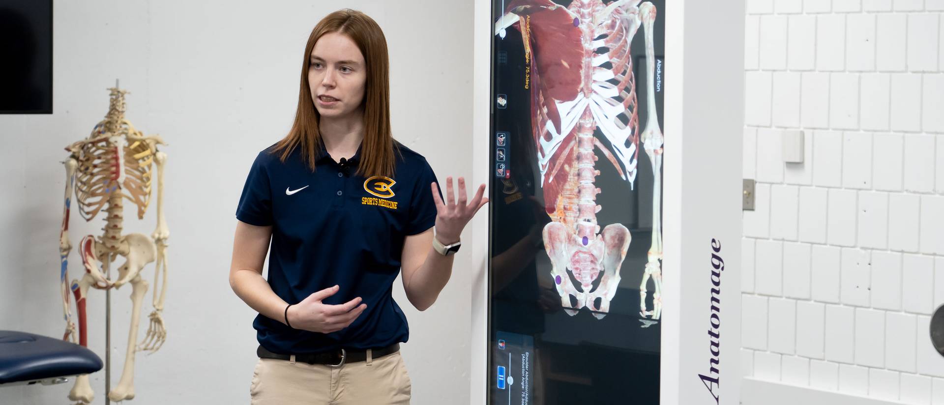
Anatomy education is coming to life for University of Wisconsin-Eau Claire students who are using new high-resolution digital cadaver tables to see the inner workings of the human body.
The College of Education and Human Sciences purchased two life-size 3D tables that are being used by undergraduate and graduate students in the departments of kinesiology and communication sciences and disorders.
“It’s such a cool piece of technology,” says Meghan Dahm, a first-year student from Hartford majoring in physical education who used the table in her anatomy course in fall 2023. “You can look at anything and everything.”
The tables are designed for use both during and outside of class to support student success, says Dr. Carmen Manning, dean of the College of Education and Human Sciences. This high-impact learning tool helps students “deeply engage with course content” in a collaborative way, Manning says.
“The Anatomage tables are one more example of how UW-Eau Claire provides excellent, engaged learning opportunities for students,” Manning says. “The anatomy learning supported by these tables is foundational to student success as they prepare for high-demand health care careers.
“Engaged, experiential learning aided by tools such as the Anatomage table are the hallmark of health sciences education at UW-Eau Claire. These tools in the hands of our talented faculty prepare students as future leaders in health care.”
The tables are used in the anatomy education course that is foundational for all kinesiology program majors and contains critical content for students to master, says Dr. Jeffrey Janot, professor and chair of the kinesiology department. Among kinesiology groups who especially will benefit from the technology are athletic training graduate students, exercise physiology graduate students and rehabilitation science students, Janot says.
Students can view and magnify any part of the human body while examining clinical scenarios through more than 1,600 case studies. The system’s software contains cases of chronic disease and conditions that impact anatomical structures in the body, so more advanced students can study those as well.
“If one can’t have actual cadaver anatomy, the images that our students are able to view and manipulate on the Anatomage table is the next best thing,” Janot says.
Tadd Turnquist, clinical assistant professor of kinesiology, implemented the anatomy tables into the curriculum in fall 2023 and plans to use them more extensively this spring semester. A dentist presenting in Turnquist’s class used the table’s touch screen to show a cadaver’s dental implants while a visiting optometrist showed different muscles of the eye on a cadaver.
“This table is one of the best tools we’ve had the opportunity to have here in kinesiology in my 14 years at UW-Eau Claire,” Turnquist says. “Students are immediately intrigued by it. They can engage with this new technology that many of them have never seen before. It’s a great way to interact and help them be engaged with the material.”
Matthew Gandrud, a first-year student from Shorewood majoring in rehabilitation science, says the anatomy table is a tremendous tool for laboratory work and preparing for exams.
“It’s been really helpful for the exams to find an exact muscle, a joint or a ligament,” Gandrud says. “It’s easier than using one of the hand models where you have to peel off muscles. This shows you the inner parts.”
Dr. Jerry Hoepner, professor of communication sciences and disorders, expects to use the digital cadaver during spring semester in multiple CSD courses, including anatomy, physiology, neurological aspects of communication and cognition, and voice and resonance disorders. CSD students will be able to use the anatomy table in small groups in conjunction with 3D models and other learning activities.
“Students will be able to look at any slice or section of the body,” Hoepner says. “It will be great for identifying muscles, as you can peel off a layer at a time to see how muscles overlap each other. It’s fantastic for examining the brain, particularly coronal, sagittal and transverse sections. Understanding these sections of the brain is often a real challenge for students.”
Story written by Gary Johnson; Video by Jesse Yang
Link to original story: https://www.uwec.edu/news/news/virtual-technology-allows-blugolds-to-use-digital-cadavers-in-anatomy-courses-5929/